Fungal Types and How They Invade a Nail
Nail fungus is not one organism. Dermatophytes are the most common group in toenails, but molds and yeasts can also be involved. The pathway matters because the organism may sit under or inside thickened nail tissue.
Common organism groups
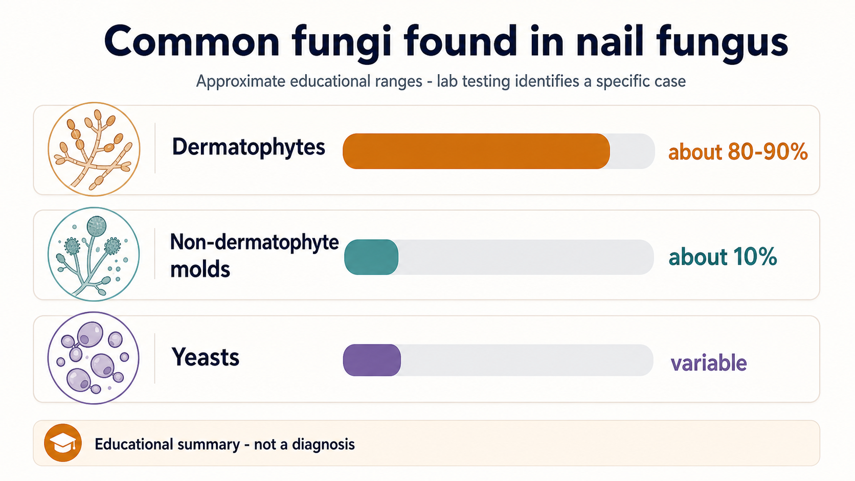
Most toenail fungal infections are associated with dermatophytes, especially keratin-loving species such as Trichophyton rubrum. Non-dermatophyte molds are a smaller but important group, and yeast involvement varies by nail type and setting.
- Dermatophytes: often described as the dominant group in toenail onychomycosis.
- Non-dermatophyte molds: globally often around 10%, with regional variation and possible mixed infection.
- Yeasts: more commonly discussed in fingernails, but can appear in nail disease.
How invasion can happen
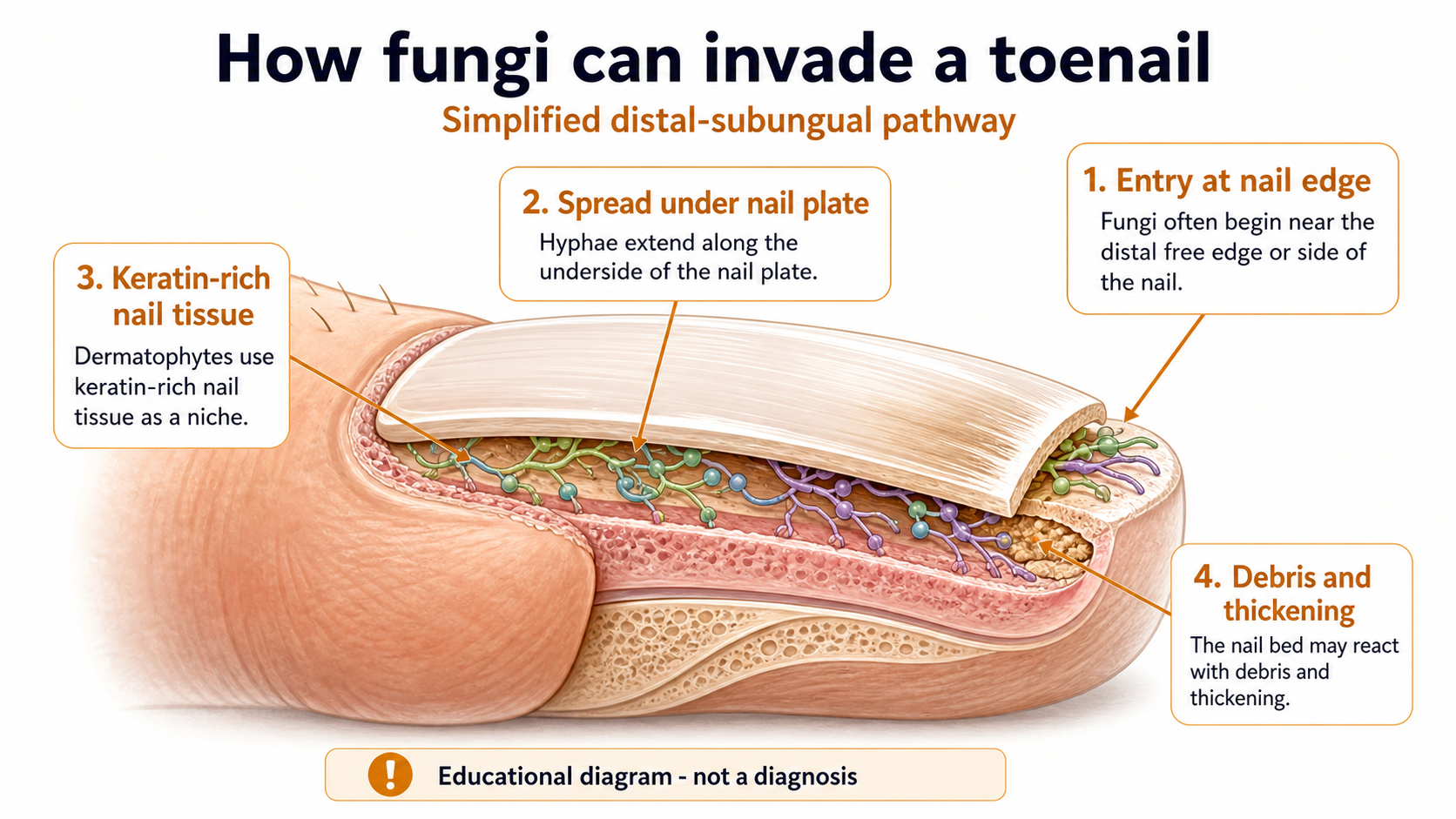
The most common pattern is often described as distal and lateral subungual onychomycosis. In plain English: the process can start near the free edge or side of the nail, then spread along the underside of the nail plate and nail bed.
As debris builds up and the nail bed reacts, the nail can look thicker, yellow, crumbly, lifted, or separated. Similar appearances can also come from trauma, psoriasis, eczema, or other conditions, so confirmation matters.
Meet the species — and their real iodine numbers
Below each cartoon is the measured log reduction for povidone-iodine from a published EN 1275 suspension test: a 4-log reduction means 99.99% of the organism was killed, the bar for calling an antiseptic fungicidal in that test. The cartoons exaggerate personality, not the data.

Red dermatophyte
By far the most common cause of toenail fungus. Slow but relentless. Log reduction 4.18 — killed.
Killed in vitro · log 4.18
Powdery dermatophyte
Second most common; often behind athlete’s foot flares. Fast-growing, heavy spore producer. Log reduction 4.29 — killed.
Killed in vitro · log 4.29
Budding yeast
More often discussed in fingernails and wet-work hands. Grows as round budding cells. Log reduction 4.39 — killed.
Killed in vitro · log 4.39
Wrinkled yeast
The most iodine-sensitive of the group in suspension tests. Log reduction 4.50 — killed.
Killed in vitro · log 4.50
Black mold
A non-dermatophyte mold — a minority of cases, but the stubborn one. Log reduction 3.10: reduced, below the 99.99% fungicidal bar.
Reduced, not killed · log 3.10Share of infections varies by region and nail type; dermatophytes dominate toenails, yeasts are discussed more in fingernails, and molds such as Aspergillus are a recognized minority. A laboratory culture is the only way to know which organism is in a given nail.
To watch these species germinate, spread, sporulate, and meet an iodine drop in motion, open the interactive Fungus Lab simulation.
What this means for home tracking
- A surface stain or color change is not enough to identify the organism.
- A thick nail can block contact between a topical liquid and the area where organisms sit.
- Improvement should be judged by new growth from the base, not by expecting the old nail to turn normal overnight.
- Diabetes, poor circulation, pain, redness, drainage, or a lifting nail should move the decision toward professional care.
Sources used for the visual summary
- DermNet: Fungal nail infections
- DermNet: Non-dermatophyte mould onychomycosis
- StatPearls / NCBI Bookshelf: Onychomycosis
- Clinical Microbiology Reviews: Pathogenesis, diagnosis, and management
- Silva-Neves V, et al. In vitro study of antiseptic solutions against onychomycosis isolates. Scientific Reports 2021;11:12789 — source of the EN 1275 log-reduction values shown on the species cards.
- Thongkham K, et al. Inactivation of dermatophyte arthroconidia. Veterinary World 2022;15:1373–1379